
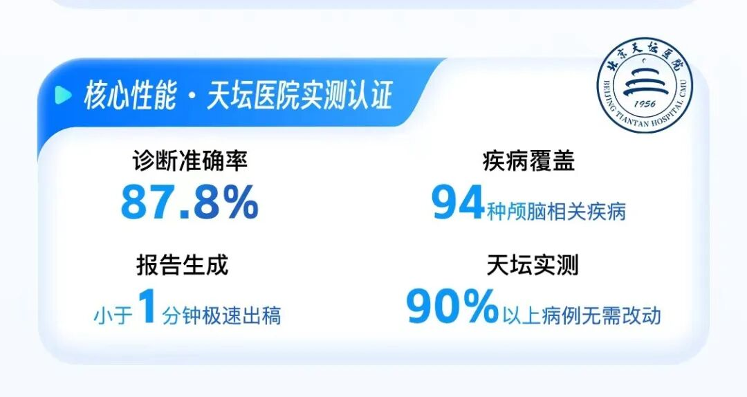
How long does it take to read a brain CT scan? For a skilled radiologist, from retrieving the images to finishing the report, it takes about 15 minutes; for complex cases, it takes at least 20 minutes. This is the work pace accumulated over years in the neuroimaging department. This time might seem a bit slow, but it is already the limit of the human brain in processing hundreds of possible lesions within a single cranial cavity. In April this year, an AI product named the Brain CT Super Agent (Dr. Xiao Jun 2.0) was launched for testing at Beijing Tiantan Hospital, Capital Medical University. It does the same thing, but generates the report in less than 1 minute—covering 94 brain-related diseases, with a primary diagnostic accuracy of 87.8%. In actual tests, over 90% of the cases could be used directly by reviewing physicians without a single word changed. The real-world data of the Brain CT Super Agent has invigorated the industry.
According to introductions, the Brain CT Super Agent (Dr. Xiao Jun 2.0) is an AI product jointly developed by Tiantan Hospital and Yinghe Yimai, an artificial intelligence enterprise incubated by Yimai Sunshine. Its emergence heralds the onset of a paradigm revolution in medical imaging AI, transitioning from a "single-disease tool" to "examination-item-level intelligent infrastructure." On April 28, 2026, this "Brain CT Super Agent" was officially brought to the forefront by Yimai Sunshine as the core product of its annual strategic upgrade. It is positioned as the "world's first examination-item-level intelligent diagnostic assistant covering all disease types," and is also endowed with broader implications and a promising future.
Frankly speaking, the medical AI industry is not short of impressive numbers. Over the past few years, products capable of obtaining Class III medical device certificates and holding new product launch events have been lining up. But an essential problem facing medical AI is: Most ordinary people are not clear why they need to pay attention to this matter.
The answer lies in the daily routine of the radiology department in any tertiary Grade A hospital. The annual growth rate of radiologists in China is only about 4%, while the growth rate of examination volume exceeds 30%. The figures at the grassroots level are even worse; some county hospital radiology departments have only two or three people, working in 24-hour shifts, and report backlog until the next day is the norm.
Brain CT happens to be one of the most frequently ordered examinations in the emergency department. For cerebral hemorrhage, cerebral infarction, and craniocerebral trauma, every extra minute of waiting means a one-minute delay in decision-making. In the reality where medical resources become increasingly scarce as they go down the hierarchy, doctors who can read brain CTs and equipment capable of performing this examination may have never been properly matched.
Therefore, when the industry talks about AI-assisted diagnosis, it corresponds not to a technical concept, but to an extremely specific supply-demand gap. It is precisely for this reason that the brain CT track has always been regarded as one of the most difficult, yet most valuable, highlands in medical imaging AI.
1. From "Drawing a Box" to "Writing a Report"
In 2024, a Boston startup named a2z Radiology AI emerged from stealth mode. The founder, Pranav Rajpurkar, is an associate professor at Harvard Medical School, specializing in general medical AI. What they do sounds uncomplicated: a single model simultaneously analyzing multiple acute conditions on abdominopelvic CTs.
The implementation results are also solid. At the end of 2025, a2z-Unified-Triage received FDA approval, becoming the first system in the U.S. market capable of simultaneously detecting 7 acute abdominopelvic conditions in a single scan. Small bowel obstruction, acute pancreatitis, and free intraperitoneal air are all on a single list.
A prospective study released at RSNA 2025 further showed that with AI assistance, reporting time was reduced by 17.8%, and the psychological burden on radiologists decreased by 22.4%.
This is the report card delivered by the abdominopelvic region, one of the tracks with the largest CT examination volume and the most complex cases. Securing this category is already impressive enough in the medical imaging AI circle.
But brain CT is on another level of difficulty.
Although there are many abdominal organs, they follow a relatively fixed screening logic to some extent: liver, gallbladder, pancreas, spleen, kidneys, and intestines. Scanning organ by organ, most acute conditions are distributed along common pathological pathways.
The brain is not like this.
A single cranial cavity is packed with brain parenchyma, cerebral blood vessels, the ventricular system, and skull structures. Lesions can range from millimeter-scale aneurysms to hemisphere-spanning space-occupying lesions, from fatal cerebral hemorrhages within minutes to degenerative changes lurking for years. Moreover, they often mimic each other on imaging; an infarction in the thalamic region and a low-grade glioma might differ by only a few grayscale points on a non-contrast CT.
The "Brain CT Super Agent" has achieved the goal of "comprehensive evaluation like a doctor" in the head, the most difficult part, and goes beyond merely marking problems to directly generating complete reports.
According to Liu Ya'ou, Director of the Department of Medical Imaging at Beijing Tiantan Hospital, the biggest threshold the "Brain CT Super Agent" has overcome, which has long plagued the industry, is that it has upgraded from image recognition to image understanding. It takes imaging input once and directly outputs a complete structured diagnostic report.
"This model has been officially put into use in our department. By opening the PACS system and clicking the Yinghe Yimai report button, the results can be automatically generated with one click. Doctors only need to review and cite them."
In Liu Ya'ou's view, this model greatly shortens the workflow of radiologists, resulting in a tremendous improvement in human efficiency.
In the medical AI industry, the birth of the "Brain CT Super Agent" means that the industry has already developed into its third stage.
The first stage is single-disease recognition, where one model identifies one disease, driven by data; the second stage is multi-disease in a single region, where one model covers multiple lesions in one region; the current third stage is complete delivery of all disease types across all anatomical structures covered by taking all examination items as units, driven by models rather than data.
The aforementioned a2z Radiology AI can be regarded as a product of the second stage. The same applies to the Chest CT Pathway-Level AI-Assisted Diagnostic Tool (AIR). It was released last October by Yinghe Yimai (also incubated by Yimai Sunshine) in collaboration with West China Hospital of Sichuan University.
AIR can perform a single non-contrast CT scan to simultaneously complete full-lesion detection and diagnosis of multiple organs such as the lungs, pleura, and mediastinum, covering all common disease types in this region.
The release of AIR was highly praised by the industry as marking the transition of medical imaging AI from the past "small model single-disease detection" (1.0 era) to the "foundation large model-driven multi-organ full-disease collaborative diagnosis" 2.0 era.
The Brain CT Super Agent has not only achieved a significant leap in the number of lesions detected—covering 94 brain-related diseases—but has also completed the functional transition from "detection" to "writing complete reports." From the output of the Chest CT AIR to the output of the Brain CT Super Agent, in just half a year, Yinghe Yimai, as the same production entity, has rapidly produced multiple AI fruits, taking the lead in pulling the entire industry into the General AGI 3.0 era of medical imaging.
2. Technological Leap as Well
But intrinsically, the biggest difference between the "Brain CT Super Agent" and previous similar medical AI technologies or products lies in its greater emphasis on the intrinsic value contributed by doctors and hospitals to the product.
At the launch event, Xu Lei, Director of the Department of Medical Imaging at Beijing Anzhen Hospital, Capital Medical University, made a vivid remark: with the help of AI, radiologists transition from report writers to AI creators, trainers, and reviewers, able to achieve experience replication and 24-hour service through digital avatars.
In fact, version 1.0 of Dr. Xiao Jun was a collaboration between Tiantan Hospital and Beijing Institute of Technology, achieving a text-to-text technological breakthrough at that time. After version 2.0 of Dr. Xiao Jun (Brain CT Super Agent) collaborated with Yinghe Yimai, Tiantan Hospital's resources actually did not change much, but Yinghe Yimai gained a say in medical AI large models.
In September last year at the Beijing CIFTIS, Yinghe Yimai, the National AI Application Pilot Base, and Beijing Tiantan Hospital jointly released a systematic R&D achievement in brain medical imaging AI: the Brain CT Core Definition Table and the Brain L1 Foundation Model.
This systematic R&D achievement and the L1 foundation model are the publicly traceable origins of the Brain CT Super Agent. The former solved "how to label data," and the latter solved "how to build the model."
As the incubation entity, Yimai Sunshine supported the generalization function of the underlying technical model with tens of millions of data points from over a hundred imaging centers. These data are not clean samples meticulously selected in a laboratory, but come from real clinical environments. After standardized labeling through a unified core definition table, they are fed back into model training.
At the technical level, Yinghe Yimai introduced two core building blocks.
The first is the self-developed "Yinghe Miaoya®" medical imaging foundation large model, an L0-level full-modality foundation model capable of processing multi-modal data such as CT, MR, and X-ray, and covering over 200 common diseases across the whole body. Via foundation pre-training plus downstream fine-tuning, the development cycle for new AI products can be shortened by more than 70%.
The second is the full-chain AI Agent matrix, responsible for breaking down diagnostic logic into multiple agents working collaboratively. The foundation provides cognitive capabilities, while the Agent matrix handles workflow orchestration. The two building blocks interlock to support the capability of single input and complete output.
With the support of the aforementioned technology and data, the Brain CT Super Agent can achieve a leading position, and more importantly, it has completed clinical implementation in the radiology department of Tiantan Hospital.
It sounds like marketing jargon, but when placing a2z's abdominopelvic achievements alongside the objective difficulty of brain CT, the weight of clinical implementation becomes evident.
In the abdominopelvic region, a2z has proven that a single model simultaneously detecting 7 acute conditions can obtain FDA approval and achieve quantifiable improvements in clinical efficiency. a2z's current product form is closer to AI-assisted report drafting, meaning the AI outputs a structured draft for physicians to review and sign.
The Brain CT Super Agent, on the other hand, has already achieved one-click report generation without modifications for over 90% of cases at Tiantan Hospital, and has successfully driven commercial implementation.
More importantly, the local iteration concept of Yimai Sunshine and Yinghe Yimai, which is to let the AI continuously evolve locally in the hospital based on the doctors' hospital-specific usage and diagnosis/treatment habits, becoming more adaptable the more it is used. Thus, a closed-loop optimization logic is formed: from doctor operation → data memory → training samples, realizing full-process data value transformation.
Gu Yun, a professor at Shanghai Jiao Tong University, describes it as: work is training, usage is evolution, making the clinical workflow the driving force for model evolution.
3. Commercialization is the Ultimate Test
If one were to find a magic mirror for the medical AI industry, it would be "whether it charges money or not."
Over the past five or six years, more than 200 AI medical devices in China have obtained NMPA Class III certificates, with 41 new ones added in 2025 alone.
But obtaining a certificate does not equal making money. The fate of a large number of products after entering hospitals is: they are installed, used occasionally, and not a single cent is collected. Hospital budgets are already tight, and AI software is not in the medical insurance catalog, so the willingness to pay is naturally discounted. The industry once fell into a vicious circle: more and more products, but no increase in revenue.
Therefore, the fact that the commercialization of the Brain CT Super Agent can truly take the lead in implementation is worth discussing separately.
Why can it charge money? The core logic is not complicated. What it solves is not optimizing experience, but increasing capacity.
Reducing the time for a brain CT report from 15 minutes to 1 minute is an efficiency boost for tertiary Grade A hospitals and a capacity supplement for grassroots institutions. When value can be quantified, the bill for paying can be justified. This forms a clear divide from those past AI products that were "quite useful but couldn't charge money."
Of course, talking about commercialization does not mean it will be smooth sailing from now on. The medical industry is inherently conservative, and doctors' trust in new tools takes time to accumulate. An unavoidable question always hangs overhead: if an AI-generated report happens to have errors, who takes the responsibility?
This is not a question that algorithms can answer; it is harder to resolve than any technical metric. The commercialization of medical AI is essentially the commercialization of trust, and money is just the final form of settlement.
From this perspective, the true meaning of the "first revenue-generating agent" is actually that someone is willing to pay for this trust. Although the scale of how much they pay has not yet been announced, the door has been pushed open a crack.
What distinguishes a product that can charge money from a product that can continuously charge money is whether it can self-evolve.
Many medical AI products encounter the same embarrassment after implementation: they become less effective the more they are used.
Diseases change, data grows, but the model is static. To update it once requires re-labeling, re-training, and re-deploying, with a cycle starting at several months. A hospital spends a sum of money, only to find a year later that the version is still the same, and naturally does not renew it the second year.
The solution provided by Yimai Sunshine and Yinghe Yimai is a three-tier architecture: data governance layer, model iteration layer, and scientific research incubation layer. These names sound a bit convoluted, but their logic becomes clear when broken down.
The bottom layer is the data governance layer.
Yimai Sunshine's national imaging center network has accumulated nearly 30 million cases and 7 PB of medical imaging data. But raw data cannot be used directly; data from different devices, different hospitals, and different periods have a wide variety of formats and labeling standards.
Yimai Sunshine's approach is to take the lead in formulating standardized specifications for data governance, and through the "Core Definition Tables" for over a dozen regions such as the brain, thorax/abdomen, and joints formulated by Yinghe Yimai, it is equivalent to setting a unified "ruler" for data labeling. Incoming data goes through governance first before moving up.
The middle layer is the model iteration layer.
The foundation model Yinghe Miaoya® is not a single-task model, but an L0-level foundation capable of processing multi-modal data such as CT, MR, and X-ray. Via foundation pre-training plus downstream fine-tuning, the new product development cycle can be shortened by more than 70%. Moreover, this model has a practical feature: accuracy can also be improved by 10%-20% under small-sample training, meaning it can quickly adapt even when there is not much labeled data for a specific disease.
The top layer is the scientific research incubation layer.
This is where the slogan "letting those who understand medicine best train the AI that understands medicine best" is truly implemented.
Top-tier institutions like Tiantan Hospital provide clinical knowledge systems and real-world feedback. Doctors are not mere tools for labeling data, but the leaders of AI R&D. This is about transforming the cognitive assets of top experts from top medical institutions into iterative fuel for AI evolution.
The operational logic of the combined three-tier architecture is that real-world data from the clinical frontline enters the governance layer, undergoes standardized processing to achieve general model upgrades, and is then led by a top-tier medical team for scientific research incubation.
The entire process does not end when the product goes to market; rather, the product launch is just the beginning. Yimai Sunshine has even set up some of its imaging centers as verification and testing bases for model adjustments and agents. Radiologists use the product in real diagnostic scenarios, discover defects, and provide feedback to Yinghe Yimai's technical team for targeted optimization.
To put it in plain language, this is not a software that is finalized upon purchase, but a digital doctor that can continuously grow through use. The capabilities of the version you use today and the version half a year later will be different.
For hospitals, this means the money spent is not a sunk cost, but an appreciating asset. This is the truly powerful aspect of self-iteration: it extends the commercial lifespan of AI products from a one-time transaction to lifelong service.
Looking back, this architecture also aligns with Yimai Sunshine's previous strategic positioning.
In its 2024 annual report, Yimai Sunshine proposed the concept of "Scenario + Data + AI Scenario," which sounded like top-level design at the time. Now, the Brain CT Super Agent has run through a complete path: massive data generated by the imaging center network, the foundation model providing the technical base, and top-tier hospitals injecting clinical wisdom, ultimately producing a product that can charge, iterate, and be reused in the brain, one of the most complex regions.
4. Epilogue
Let's turn the clock back to September 2025, at the AI + Healthcare Seminar of the CIFTIS.
That launch was not very high-profile in terms of volume. What was showcased on stage were two somewhat dry-sounding things: the Brain CT Core Definition Table and the Brain L1 Foundation Model.
The former unified the labeling specifications and structured report standards for brain imaging, solving the problem of data from different hospitals, different devices, and different periods speaking different languages; the latter was iterated on the self-developed Yinghe Miaoya L0 full-modality foundation by Yinghe Yimai, specifically optimized for the brain.
These two seemingly unglamorous outputs answered a question that few people publicly discuss: how exactly do the data and knowledge from top-tier hospitals become "nutrients" that AI can digest?
Simply put, the core definition table sets the standards, and the L1 model builds the skeleton. Seven months later, this set of standards and skeleton grew into the Brain CT Super Agent in the real-world scenarios of Tiantan Hospital.
Thus, China's medical imaging AI has crossed a watershed.
In the past, what AI did was very simple: circle the suspicious areas and leave the rest to the doctors. This step was hard, but it was still a matter of perceptual assistance. Now, this product has moved from helping you see to thinking about how to write it for you, taking a step into the realm of cognitive generation.
This is not just a few percentage points jump in accuracy; the dimension of capability has changed.
This is precisely the Medical AI 3.0 era defined by Yimai Sunshine and Yinghe Yimai: taking examination items as units, batch-developing AI tools, full-modality, full-examination, full-process, fully automated, truly embedded into the workflow of medical technicians and nurses, and using models to drive complete delivery. The pragmatic form of AGI in the medical imaging field is not an omnipotent doctor from a sci-fi movie, but an agent with comprehensive diagnostic capabilities close to those of human doctors on complete examination items. The "Brain CT Super Agent" is the first product to turn this definition into reality.
Looking at a deeper level, its significance is not just that a product is powerful, but that the entire paradigm behind it has been run through from end to end.
The past medical AI R&D paradigm was project-based: one hospital, one project, one disease, one set of data, one model. Every time the scenario changed, it had to start over, resulting in long R&D cycles, high replication costs, and difficult commercial translation.
And now, this new paradigm is an agent factory of foundation model + expert co-construction + data closed-loop: the imaging center network provides data from real scenarios, top-tier hospitals inject clinical cognition into the model, and the foundation drives continuous product iteration.
This kind of thing used to appear more in technical white papers, but now it has been run through from beginning to end by a single product line.
This is a paradigm shift in the development of China's AI healthcare, switching from a project-driven workshop to a foundation-driven agent factory, and from papers and demos to billing statements and clinical workflows.
If the paradigm shift only stays in theory, it is just a vision; once it appears on a hospital's bill, it becomes an industrial fact.
This industry has never lacked stories. What it lacks are products that can appear on billing statements, models that can continuously evolve, and doctors who can confidently hand over initial reports to AI. Yimai Sunshine, Yinghe Yimai, and Tiantan Hospital have gathered all these this time.
Having stepped firmly on the stepping stone of the brain, what it has stepped out is not a small path, but an entrance to an era.
Disclaimer: This article is written based on publicly available information or information provided by interviewees, but Decode and the author do not guarantee the completeness and accuracy of such information and materials. Under no circumstances shall the information or opinions expressed in this article constitute investment advice for anyone.